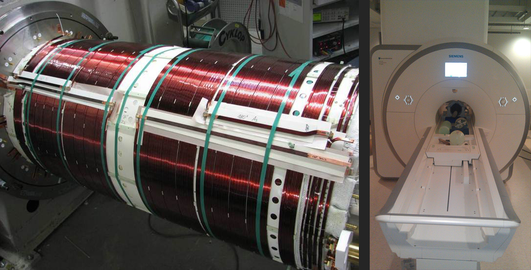
Connectome scanner installation and tune-up
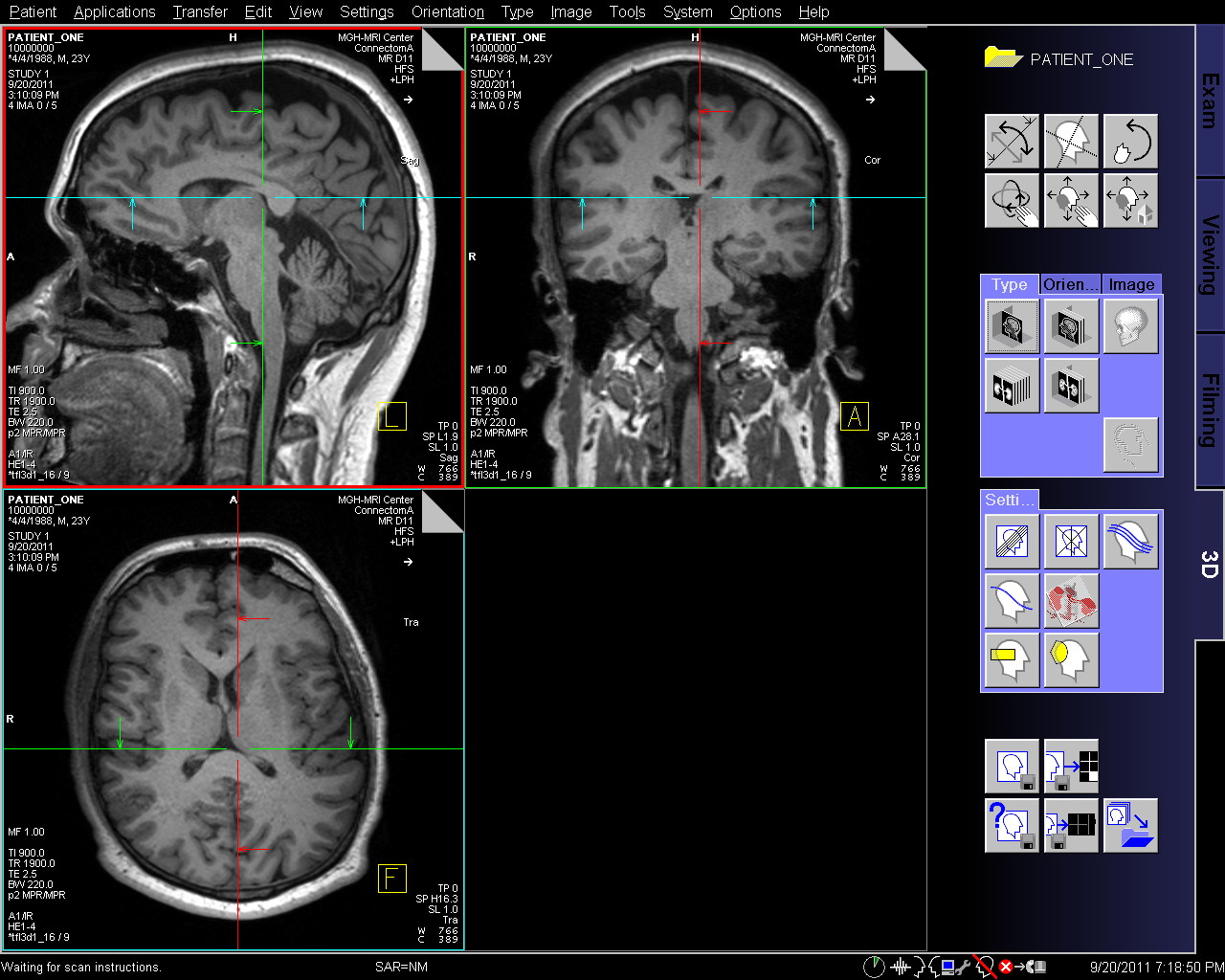
The Connectome scanner has been delivered to MGH and was officially installed and tuned up on September 20, 2011. While the system is under Siemens control, human subject testing is performed under the union of restrictions set by the MGH HRC (IRB) and Siemen’s internal testing requirements.
Though the installation and tuneup have been completed, it was not turn-key, as was expected with such a complex installation. A problem occurred with the tuning delays in the 12 gradient waveforms which drive the three gradient axis (X,Y,Z). A unique delay is calculated for each of the gradients and the Connectome scanner has four amplifiers and cable sets for each axis. This made the tuneup process significantly more involved.
Despite causing a slight time delay, addressing this challenge revealed a need for an automated procedure to conduct effective routine tuneups as well as auto adjustments when a singular component is replaced. Thomas Witzel is developing a sequence that can automatically perform this search over this parameter space, updating the delays and ultimately achieving a more streamlined tuneup.
Non-scanner related progress:
The blipped-CAIPI simultaneous multislice method needs to be tested for compatibility with the scanner to prevent 100s of GBs of data from being transferred offline for processing. This method will allow for a wide range of studies to utilize the technology and is being developed by Steven Cauley, Ph.D. and Kawin Setsompop, Ph.D. The code can be used to fit rectangular kernels across odd and even K-space lines as well as tease apart additional collapsed data sets. The integration of the Slice-Grappa code into the ICE framework is on-going.
Also in development are metrics to determine the effectiveness of a given methodology (such as the Connectome gradient, or a q-space/k-space tradeoff) in uncovering the structural connectivity in complex white matter regions. Software is being created to allow Contrast-to-Noise metrics on the ODF, as well as provide flexible evaluation of different q-space sampling strategies. A flexible tool-kit for evaluating the PDF and ODF from DSI data and assessing filtering and q-space sampling strategies is also in development.
Another area of interest is the kspace/qspace tradeoff in structural connectomics by way of acquiring and analyzing high resolution diffusion imaging. Many papers show interesting results, but none that reflect on tractography. A set of 32 channel brain array coils was developed for pediatric imaging and recent investigations have shown that it is also
suitable for adult women. This allows for a 2X SNR advantage of the conventional adult 32 channel coil. The first coil will
be used along with the 64 channel coil which has been designed to fit 98 percent of adult heads.
Analysis of brain architecture
We are continuing to investigate methods to create local coordinate systems for the brain from crossing fibers. Previously, fiber orientations were defined in terms of the extrinsic image coordinates (X,Y,Z). Now, working one group of fibers at a time, we compute the distribution of orientations for this group, reduce it to 3 principal axes, and then take these as a coordinate basis. This approach produces beautiful local 2D coordinate patches throughout most or all of the brain. Our present effort is to standardize this and devise methodology to glue together local coordinate patches into larger 3D and regional coordinates, with the ultimate aim to create a standard, unified fiber-based coordinate system for the brain.


